
Figure 2.
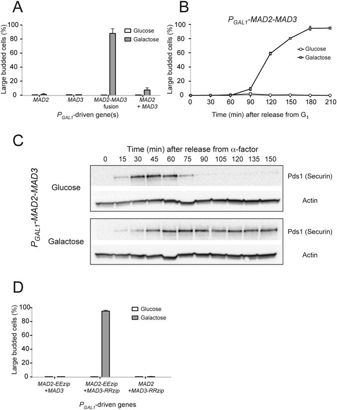
Overexpressing a Mad2-Mad3 protein fusion leads to metaphase arrest. (A) Cells with the indicated PGAL1-driven genes were grown to mid-log phase, arrested in G1 with α-factor, and were released into media with either glucose or galactose. After 3 hours of growth, the percentage of large budded cells was determined by light microscopy as a measure of metaphase arrest. Error bars represent the standard deviation of three independent trials. Two hundred cells were counted for each trial. (B) PGAL1-MAD2-MAD3 cells were released from G1 arrest into glucose- or galactose-containing media. Samples were collected at the indicated time point and the percentage of large budded cells was determined by light microscopy. Error bars represent the standard deviation of three independent trials. Two hundred cells were counted at each time point for each trial. (C) Cell cycle progression of PGAL1-MAD2-MAD3 cells was monitored by Western blots (n=3), which detect securin (Pds1), a protein that is destroyed as cells enter anaphase. Cells were grown to mid-log phase and arrested in G1 with α-factor, and were released into media with either glucose (top) or galactose (bottom). Western blots against Myc (to visualize Myc-tagged securin) or actin (loading control) were performed. When the cells were grown in glucose, securin level first increased and then dropped rapidly as cells progressed into anaphase. When the Mad2-Mad3 fusion was overexpressed in the presence of galactose, securin was stabilized, indicating that the cells were arrested in metaphase. (D) Overexpressing Mad2 and Mad3 linked by leucine zippers also induces metaphase arrest. Cells with the indicated PGAL1-driven genes were released from G1 arrest into glucose- or galactose-containing media. The percentage of large budded cells was determined by light microscopy after 3 hours of growth. Error bars represent the standard deviation of three independent trials. Two hundred cells were counted for each trial.