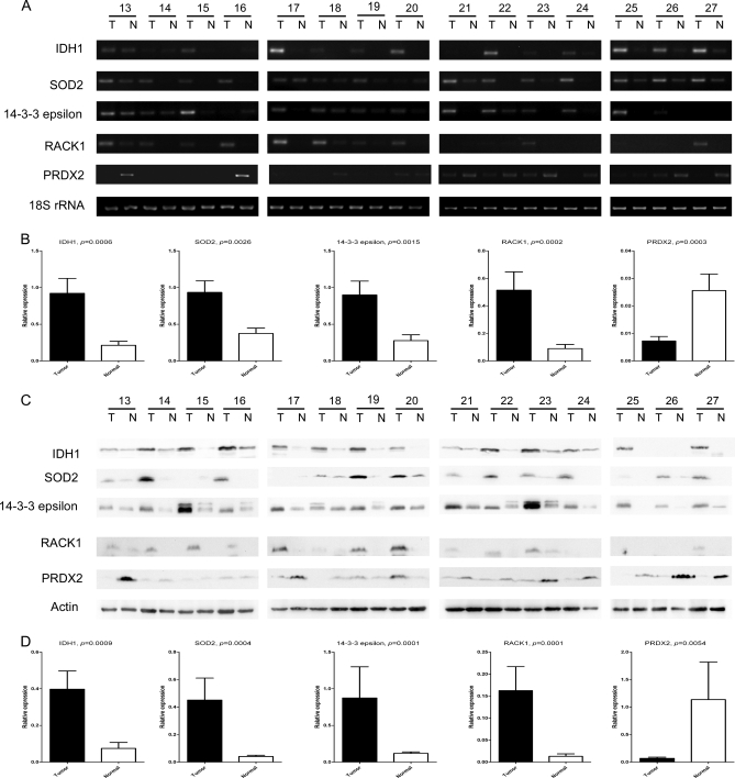
Fig. 2.
Validation of differential expression of IDH1, SOD2, 14-3-3ε, RACK1, and PRDX2 in paired lung SCC tissues by semi-quantitative RT-PCR and Western blot. Semi-quantitative RT-PCR (A) and Western blot (C) were performed in 15 independent pairs of lung SCC tumors and corresponding normal tissues. 18 S RNA and β-actin were used as internal controls. The agarose gel images (B) and Western blot images (D) were quantified by densitometric scanning, and a Wilcoxon matched pairs test was used after the intensity values were normalized against those of controls.

