Fig. 3.
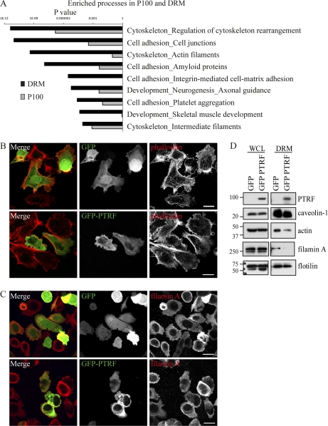
PTRF expression alters the DRM-associated pool of actin cytoskeletal proteins. A, Bar chart of GeneGo analysis displaying the most highly enriched biological processes for the P100 and DRM fraction. Cells were stained with phalloidin (B) or antibody against filamin A (C). Localization was analyzed by fluorescent confocal microscopy. No obvious change in distribution was observed between GFP and GFP-PTRF expressing cells. Scale bar = 20 μm. D, Whole cell lysates or DRMs were isolated and analyzed by Western blotting. Blots were probed with antibody against PTRF to show transfection, caveolin-1, β-actin, filamin A, and flotillin. The whole cells lysates showed no difference in protein levels between GFP and GFP-PTRF cells however, the DRM fraction showed a reduction in β-actin and filamin A in GFP-PTRF cells.

