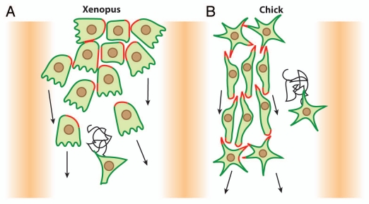
Figure 2.
Migration of cephalic NC cells in Xenopus and chick embryos. (A) In Xenopus, NC cell migration start as a loose cell sheet (top part) and progressively turns into a cell streaming composed of mesenchymal cells (bottom part). (B) In chick, NC cells migrate as mesenchymal cells and form chains. In both models cells are polarized by interactions with other NC cells (red) and maintained as a dense group by the presence of inhibitors defining the borders of the NC routes (shades of orange). High cell density leads to directional movement while isolated cells exhibit poor directionality (sinuous path).

