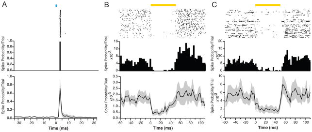
Figure 2. Optogenetic tools for activation and suppression in vivo.
A. Raster plot (upper panel) and PSTH (middle panel) of the light-evoked activity of an FS-PV+, ChR2-expressing interneuron in layer 2/3 of visual cortex in a Parvalbumin-Cre mouse after injection of AAV-DIO-ChR2-mCherry. This cell produced one spike in response to each light pulse (1ms duration) with a latency of ~3.5ms and a reliability of ~100%. A population average (lower panel) shows a high degree of repeatability of this type of response (n = 62 cells). Black line indicates mean response, gray shading indicates SEM. All data in this and other figures were recorded using custom-built dense tetrode arrays and an optical fiber. B. Raster plot (upper panel) and PSTH (middle panel) of the activity of an example RS, eNpHR v3.0-expressing excitatory neuron in layer 2/3 of visual cortex in an Emx1-Cre mouse after injection of AAV-DIO-eNpHRv3.0-eYFP. On each trial, the neuron’s spiking was suppressed in response to a pulse of yellow light. The PSTH also shows a small increase in firing immediately after the light stimulus ended. Across the population of recorded neurons (n = 34, lower panel), eNpHR v3.0 activation eliminated ~65% of spikes, but demonstrated a slow decrease in efficacy during the light pulse. C. Raster plot (upper panel) and PSTH (middle panel) of an example RS, Arch-expressing excitatory neuron in layer 2/3 of visual cortex in an Emx1-Cre mouse after injection of AAV-FLEX-Arch-eGFP. On each trial, the neuron’s spiking was suppressed during the light pulse. Across the population of recorded neurons (n = 28, lower panel), Arch activation eliminated ~70% of spikes. PSTH bins are 2.5ms.

