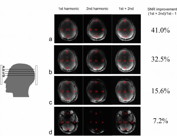
Fig. 7.
Combined human brain images with first and second harmonic coil elements and all coil elements at axial planes from (a) to (d). Ave raged regional SNR measurements are shown in the right column. The percentage of SNR improvement represents the SNR contribution from second harmonic coil elements. More SNR contribution comes from second harmonic coil elements in slices closer to the top of the human brain.

