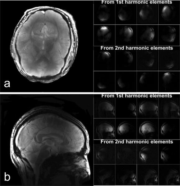
Fig. 9.
GRE data set acquired with the GE 7T scanner. (a) shows combined GRE images and sub-images from each coil element in axial plane with 20° flip Angle, TE/TR 6.9ms/100ms, 24cm × 24cm FOV, 256 × 256 matrix, 3mm slice thickness, NEX 1. (b) shows combined images acquired using the same GRE sequence but with 4 NEX and corrected signal intensity.

