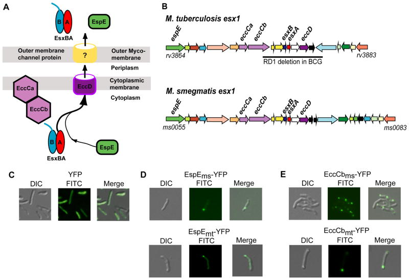
Fig. 1.
M. smegmatis and M. tuberculosis Esx-1 proteins localize to a cell pole in M. smegmatis cells. (A) Model of the ESX-1 secretion apparatus identifying key proteins discussed in this work. EccCab is an ATPase thought to deliver the EsxA and EsxB heterodimer, and associated proteins, to the pore (EccD). The pore components of the outer membrane are unknown. For simplicity, only one (EspE) of the many proteins co-secreted with EsxAB is shown. (B) Comparative genetic map of the esx1 operons of M. smegmatis and Mtb, highlighting key genes. Gene orthologs and paralogs are colour coded. The bar below the Mtb map indicates the deletion found in M. bovis(BCG) resulting in its attenuation. (C–E) Differential interference contrast (DIC, left panel), FITC (center), and merged (right) images of M. smegmatis showing that YFP is distributed throughout the cells (C), while YFP-tagged EspEms or EspEmt (D), and the ESX-1-associated ATPase, EccCbms, and it’s Mtb orthologue, EccCbmt (E), localize to the polar regions of M. smegmatis cells. 630X total magnification.

