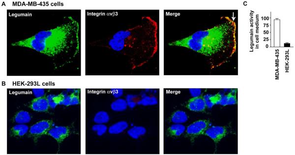
Fig. 2. Legumain is exclusively colocalized with integrin αvβ3 on the MDA-MB-435 cancer cell surface.
(A) Legumain (green) is detected in intracellular vesicles and prominently co-localized with integrin αvβ3 (red) under hypoxia. Cell nuclei are stained with DAPI (blue). Double staining is performed with anti-integrin antibodies and anti-legumain antibody. Stained cells are imaged by confocal microscopy, and the slice closest to the coverslip is presented for each cell. Note that legumain is localized on MDA-MB-435 cell surface (green, white arrow). (B) High expression legumain is only detected in intracellular vesicles of human HEK-293L cells. HEK-293 cells do not express legumain and integrin αvβ3 on cell surface. (C) MDA-MB-435 cells secret more active legumain into the culture medium than human HEK-293L does. The experiments were repeated three times (p<0.001).

