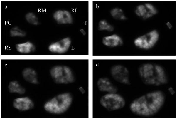
Figure 6.
Mice were exposed to an aerosol of AlPCS and the above figure of the lung lobes were obtained by converting the raw images to a 12 bit gray scale and increasing the contrast of each image in an identical manner where (a) initial excised lobes, (b) lobes after compression, (c) lobes compressed to a thickness of 0.1 cm, (d) compressed to a thickness of 0.072 cm. The lobe designations are L:left, RS:right superior, RM:right medial, RI:right inferior, PC:post caudal, and T:trachea

