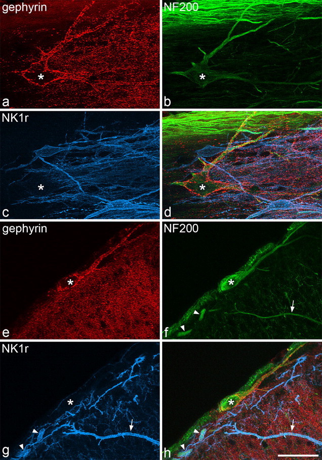
Figure 1.

Immunostaining for gephyrin, NF200, and the NK1 receptor in horizontal and transverse sections. a–d, Confocal images through part of a large lamina I cell (asterisk) in a horizontal section, scanned to reveal gephyrin (red), NF200 (green), and NK1r (blue), together with a merged image. The soma and dendrites of this neuron are coated with gephyrin puncta but are not NK1r immunoreactive. The cell contains NF200 immunoreactivity, which is relatively weak in the cell body and strong in the dendrites. Numerous axons with strong NF200 immunoreactivity are seen in the white matter in the upper part of the field. e–h, Confocal images scanned through a large lamina I cell (asterisk) in a transverse section. Again, the cell body and dendrites are NF200 positive, NK1r negative and have numerous gephyrin puncta on them. Two NK1r-immunoreactive dendrites that also contain NF200 (arrowheads), belong to a lamina I neuron, although the soma of this cell is not present in this field. The arrow shows the dendrite of a neuron from the deeper part of the dorsal horn that is immunoreactive for both NK1r and NF200. Images are made from projections of 26 (a–d) or 18 (e–h) optical sections at 1 μm z-spacing. Scale bar, 50 μm.
