Figure 3.

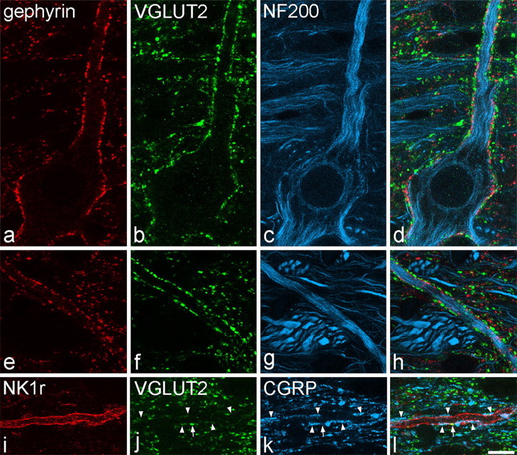
Contacts between VGLUT2-immunoreactive axons and lamina I cells in horizontal sections. a–d show confocal images scanned to reveal gephyrin (red), VGLUT2 (green), and NF200 (blue) through the cell body and a proximal dendrite of a large lamina I neuron. The cell, which contains NF200, is coated with gephyrin puncta and receives numerous contacts from boutons with strong VGLUT2 immunoreactivity. e–h show part of a dendrite belonging to this cell that is located >150 μm from the soma, scanned to reveal the same types of immunoreactivity. Again, the dendrite has numerous gephyrin puncta and is outlined by numerous contacts from VGLUT2-positive boutons. i–l, Confocal scans (NK1r, red; VGLUT2, green; CGRP, blue) through a distal dendrite belonging to an NK1r-positive lamina I cell. The dendrite receives a single contact from a bouton with strong VGLUT2 immunoreactivity (arrow) and several contacts from CGRP-immunoreactive boutons (arrowheads), some of which show weak VGLUT2 labeling. Images are projections from two (a–d) or three (e–h, i–l) optical sections at 0.5 μm z-separation. Scale bars, 10 μm.
