
Figure 1. MYXV treatment of normal and leukemic HSPCs in vitro.
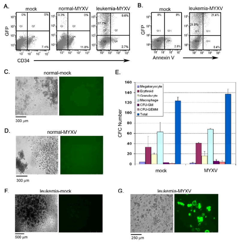
(A) Normal and leukemic cells (FLT ITD+) were incubated with MYXV expressing GFP at 10 MOI. Flow cytometric analysis demonstrated that a small population of normal MNCs was infected while a significant proportion of AML cells were susceptible to MYXV infection. (B) Analysis of apoptotic events was examined and showed that cell death was mainly observed in the GFP+ MYXV infected cell population as shown by Annexin V staining. All gating was established using appropriate isotype controls. (C, D) Normal BM cells were incubated with MYXV expressing GFP at 10 MOI and assessed for colony forming potential. Various types of CFC colonies were formed and showed no GFP expression in either MYXV-treated (D) or mock-treated samples (C) indicating that MYXV was unable to infect normal CFCs. (E) The frequency of each colony type was enumerated following MYXV or mock treatment. Similar frequencies were observed between both cohorts suggesting that MYXV does not prevent normal CFC colony differentiation in vitro. Data are representative of repeat experiments and values represent mean ± standard deviation. (F) Mock-treated AML cells formed AML-CFU colonies. (G) MYXV-treated AML cells showed signs of infection (GFP+) and did not form colonies.