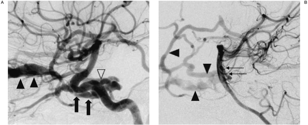
Figure 2.
A) Right internal carotid arteriogram, the cavernous sinus (arrows) and dilated SOV (full arrowheads) are seen in a lateral view, revealing the origin of the PTA (empty arrowhead). B) Left vertebral arteriogram, lateral view, showing the cisternal portion of the PTA (arrows) filling the fistula, indicating the PTA-cavernous sinus fistula. The cavernous sinus and dilated cortical veins (arrowheads) are also shown.

