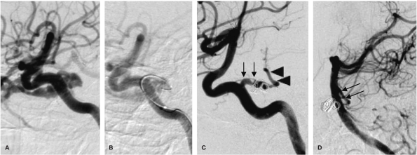
Figure 3.
A,B) Lateral oblique view during transarterial embolization. B) A microcatheter was inserted into the fistula, and the two GDC vortex coils were deployed to occlude the fistula. C) Right internal carotid arteriogram after coiling reveals a complete occlusion of the fistula. There is a connection between the preserved PTA (arrows) and the basilar artery (arrowheads). D) The left vertebral arteriogram shows filling of the PTA (arrows).

