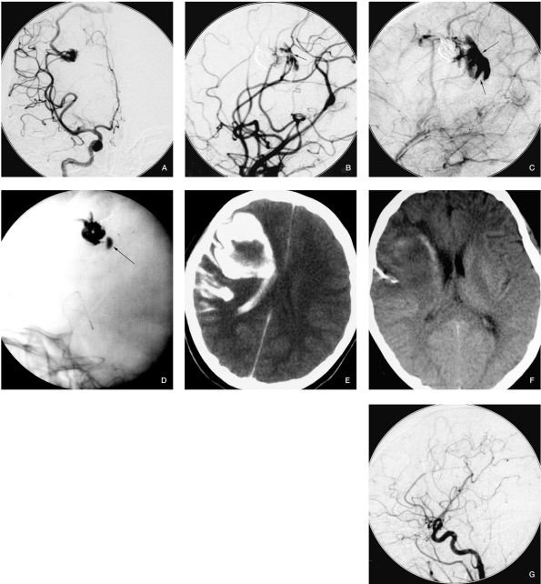
Figure 2.
Case 2. A) Angiogram before embolization. B) Contrast agent extravasation after occluding 1 feeding pedicle. C) The draining vein cannot be seen even in venous phase, and the contrast agent stagnation in the hematoma confirms draining vein occlusion. D) Contrast agent stagnation during fluoroscopy. E) CT scan immediately after embolization. F) CT scan when the patient was discharged. G) Follow-up angiography six months later, shows completely occlusion of the AVM.

