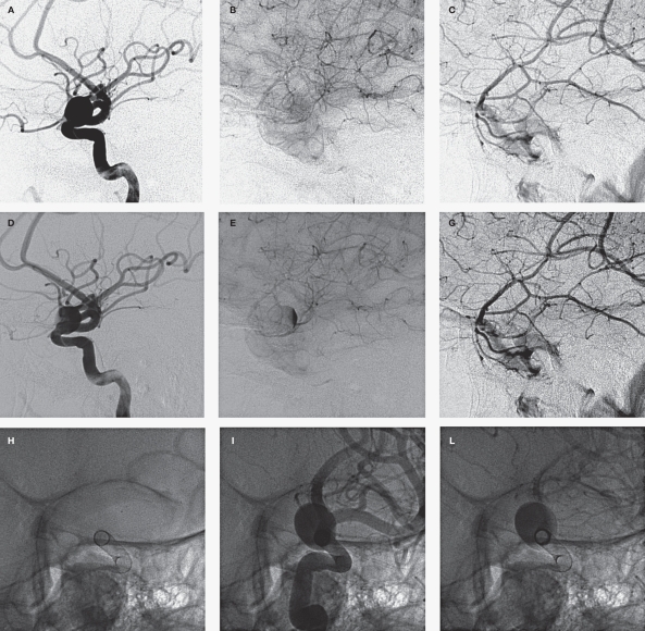
Figure 2.
Selected images of a wide-necked paraophthalmic aneurysm treated with a single flow diverting stent (Pipeline, eV3). The top panel shows the pre-treatment angiogram in the arterial (A), capillary (B) and venous (C) phases. Contrast clears from the aneurysm by the capillary phase (Grade A1). On the post-treatment angiogram (middle panel), the aneurysm continues to fill completely in the arterial phase (D), however contrast lingers into the capillary phase (E), and clears by the venous phase (F) (Grade A2). The subtle stasis is often best appreciated on an oblique projection in line with the long axis of the stent at the level of the aneurysm neck (G). The aneurysm fills completely in the arterial phase (I) but contrast clears from the vessel lumen prior to the aneurysm sac (J).

