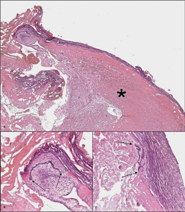
Figure 4.
Microscopic overview (A) of a part of the endoluminal thrombus (asterisk) covered by bundles of collagen fibers (Elastica-van Gieson stain, x50). Focal remnants of an arterial wall are seen (B and C). The elastic lamina (black) stops at the arrows where a pad of intimal thickening is seen (Elastica-van Gieson stain, x200).

