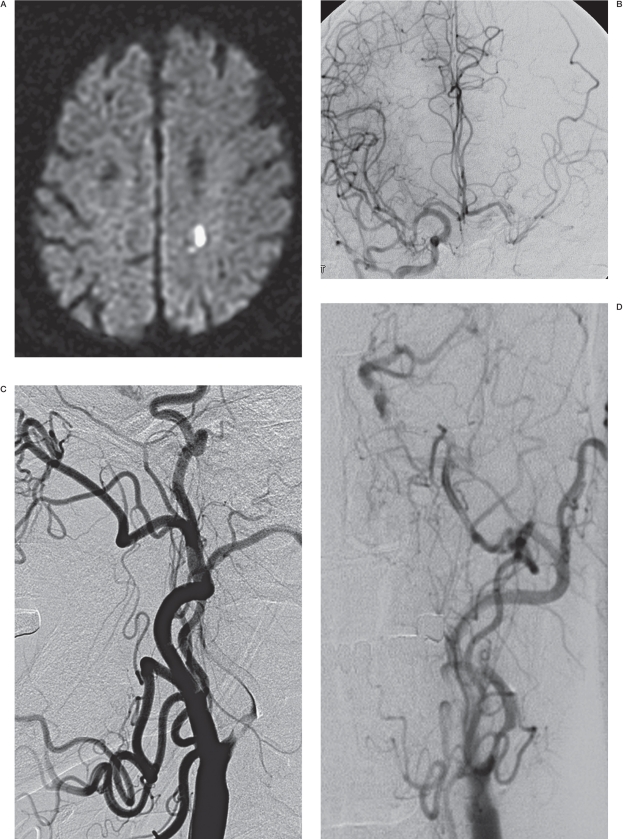
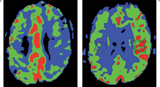
Figure 1.
A 77-year-old male presenting with aphasia (NIHSS = 3). A) Diffusion-weighted image showing an acute focal ischemic change in the left corona radiata. B) Right carotid angiogram showing incomplete cross filling through the anterior communicating artery. C, D) Left carotid arteriograms. C) Anteroposterior view, showing partial filling of the left middle cerebral arteries via collaterals through the external carotid artery. D) Lateral view, showing near occlusion of the left carotid bulb with a stasis of contrast agent in the left cervical internal carotid artery. Following successful stenting, this patient experienced a sudden deterioration of his neurological status, with no evidence of hemorrhage or further infarction on diffusion-weighted images (not shown). E) MR perfusion study showing decreased blood flow in the left MCA territory and revealing diffusion and perfusion mismatch corresponding to patient symptoms before stenting. F) MR perfusion performed immediately after stenting, showing hyperperfusion in the left brain. This patient recovered after strict blood pressure control and experienced no adverse events during the 16-month follow-up period.


