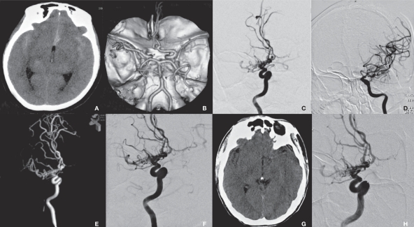
Figure 1.
Radiological imaging of MMS associated with the anterior communicating artery aneurysm in the saccular group (case 1). A) CT shows SAH. B) CTA shows an anterior communicating artery aneurysm and right MMS. C,D) DSA shows an anterior communicating artery aneurysm and right-side MMS. E) 3D-DSA provides an optimal view of aneurysm embolization. F) DSA shows a well-developed left carotid artery (arrow). G) Repeated DSA shows no recurrence of aneurysm (arrow).

