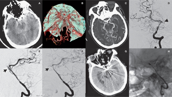
Figure 2.
Radiological imaging of MMD associated with basilar tip aneurysm in the saccular aneurysm group (case 7). A) SAH is shown. B) CTA shows MMD and basilar tip aneurysm. C,D) DSA shows the aneurysm (arrow) and extended blood supply from posterior circulation (ellipse). E) DSA shows basilar tip aneurysm (arrow). E) aneurysm embolization is shown. F) Repeated CT scan six months after surgery reveals radial artifacts. G) Repeated DSA shows no recurrence of aneurysm.

