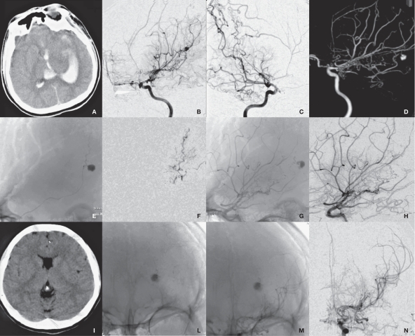
Figure 3.
Radiological imaging of MMD associated with left anterior choroidal artery aneurysm in the pseudoaneurysm group (case 1). A) CT shows IVH. B-D) DSA shows a anterior choroidal artery aneurysm and MMD. E) Microcatheter superselective angiography. F,G) DSA shows the parent artery and the embolized aneurysm. H-I) Repeated DSA shows no recurrence of aneurysm.

