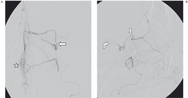
Figure 2.
SA. Frontal (A) and lateral (B) views of selective injection into the left sphenopalatine artery, one day after previous embolization, exhibit some nasal blush (asterisk), as well as collateral flow into the left ophthalmic artery (straight arrows). There is also mild washout of contrast into the left ICA on the lateral view (curved arrow).

