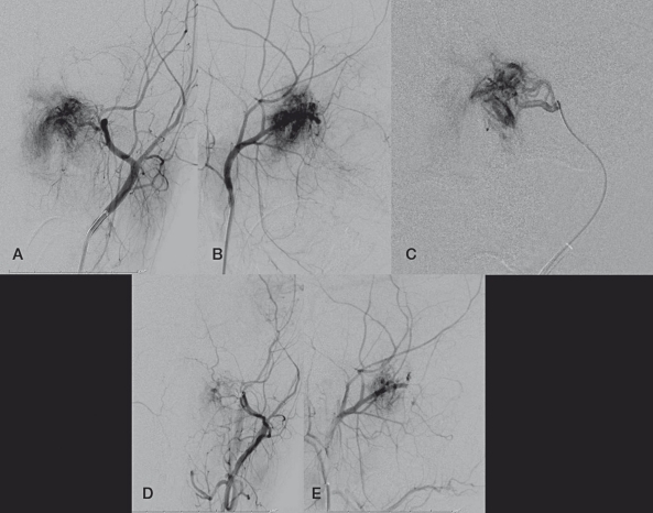
Figure 1.
A, B) Frontal and lateral views of a left external carotid artery injection demonstrating the hypervascular nasopharyngeal JNA. C) Frontal view of a microcatheter injection of the left internal maxillary artery. D,E) Frontal and lateral views of the left external carotid artery after the embolization, demonstrating devascularization of the mass.

