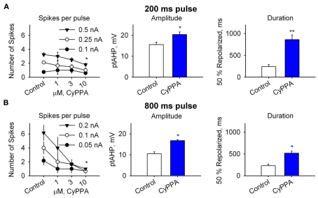
Figure 3.
Summary of the effects of current pulse intensity [(A): 200 ms; 0.1, 0.25, 0.5 nA; (B): 800 ms; 0.05, 0.1, 0.2 nA] and CyPPA concentrations (1, 3, and 10 μM) on DA neuron activity, ptAHP-amplitude, and -duration. The number of spikes decrease concentration-dependently most obviously at the longer pulses, reflecting the significance of increasing mAHP (n = 8, 3, 4, 5, 200 ms pulse; n = 6, 3, 3, 3, 800 ms pulse). Middle panels, peak amplitude of ptAHP compared with baseline membrane potential (Vm) before and after 10 μM CyPPA (n = 3). Right panels, time for 50% repolarization from peak of ptAHP to baseline Vm before and after 10 μM CyPPA (n = 3). *P < 0.05 and **P < 0.01.

