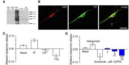
Figure 5.
CyPPA inhibits spontaneous and evoked release of dopamine form TH/SK3 positive cultured rat midbrain neurons. (A) Western blot detection of SK3 in rat midbrain cultures. Extracts of HEK293 cells transiently transfected with rSK1, rSK2, and rSK3 and adult rat brain membranes (RBM) as well as lysates from cultured midbrain neurons were separated on 4–15% SDS-PAGE and immunoblotted with anti-SK3 antibody. A band of approximately 70 kDa was detected in midbrain cultures, RBM lysates, and HEK293 cells transfected with rSK3. This band was absent in lanes loaded with HEK293 cells expressing rSK1 and rSK2. (B) Immunodetection of SK3 subunits in rat midbrain cultures. Cultured midbrain neurons (8 days in vitro) were double-labeled for SK3 and tyrosine hydroxylase (TH). In TH-positive dopaminergic neurons, the SK3 subunit displayed a primarily somatic–dendritic localization whereas little immunoreactivity was associated with axons (arrow). Scale bar 20 μm. (C) Basal release and stimulated release with 25 mM KCl or inhibited release with 30 μM Cd2+ and with 1 μM TTX. (D) Basal release and stimulated release with 3 μM haloperidol and inhibited release with 10 μM quinpirole. Blue bars represent inhibited release with increasing concentrations of CyPPA (*P < 0.05, **P < 0.01). The data presented are representative of three independent experiments.

