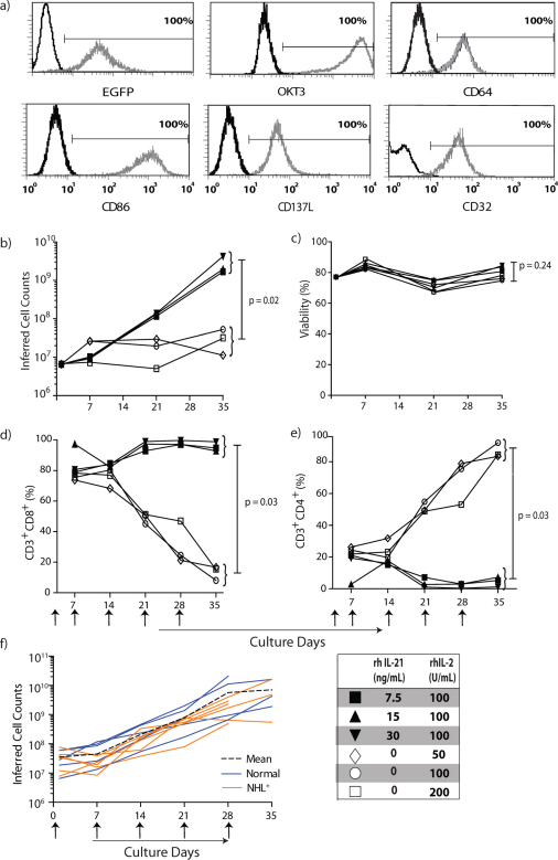
Figure 2. rhIL-21 preferentially propagates canine CD8+ T cells on OKT3-loaded aAPC.
T cells isolated from PBMC of healthy dogs were numerically expanded on γ-irradiated OKT3-loaded aAPC at a T-cell : aAPC ratio of 2:1. T cells were re-stimulated with aAPC every 7 days. Recombinant human IL-21 and/or rhIL-2 were added to the co-culture of T cells on aAPC as shown. (a) Parental K562 cells were genetically modified to co-express human CD64 and the human T-cell co-stimulatory molecules CD86, CD137L, and membrane-bound IL-15 (co-expressed with EGFP) and cloned (CLN4). OKT3 was loaded via introduced CD64 on CLN4 and detected with a Fab-specific antibody. CLN4 is shown as gray-open histograms and K562 parental cells as black-open histograms. (b) Numeric expansion of T cells on γ-irradiated OKT3-loaded aAPC (added every 7 days) in the presence of rhIL-2, or both rhIL-2 and rhIL-21. The average cell counts from two donors are shown. (c) The mean viability of T cells during co-culture on aAPC and cytokine(s) (n = 2). Percentage expression (n = 2) of (d) CD3+CD8+ and (e) CD3+CD4+ T cells upon co-culture with aAPC and cytokines(s). (f) Numeric expansion of T cells measured over culture time on aAPC and rhIL-2 and 21 recorded as the mean inferred cell count (black dashed line) based on 5 healthy subjects (blue solid line) and 5 non-trial canines with NHL (orange solid line). Upward arrows indicate timing for the addition of γ-irradiated OKT3-loaded aAPC.

