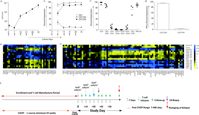
Figure 3. Characterization of T-cell infusion products.
(a) The mean inferred cell count for 6 (of the 8) canines infused with T cells that were propagated over 28–35 days on γ-irradiated OKT3-loaded aAPC (CLN4) in presence of rhIL-2/IL-21. Arrows represent days aAPC were added. (b) Percent expression of CD4+ and CD8+ T-cell subsets during propagation on γ-irradiated aAPC in the presence of rhIL-2/IL-21. Arrows represent days aAPC were added. (c) Immunophenotype of the T-cell infusion products (n = 6) at the time of cryopreservation. The horizontal lines describe mean percentage expression. (d) T-cell infusion products were tested for IFN-γ production after stimulation with OKT3-loaded aAPC in the presence of rhIL-2/IL-21 (n = 3). Analysis of multiplexed digital gene profiling of PB-derived canine T cells before and after propagation from healthy subjects (n = 2) and canines with NHL (n = 6) identified mRNA species which were either significantly (p<0.001) (e) up-regulated (> 2 fold change) or (f) down-regulated (< 2 fold change) in both healthy subjects and in at least 5 of 6 canines with NHL. (g) Study design: Enrollment occurred pre-, post, or during treatment with CHOP. Enrolled canines received one course of CHOP, typically administered over 19 weeks, and received one to three infusions of T cells 14 days apart using an intra-patient dose-escalation scheme.

