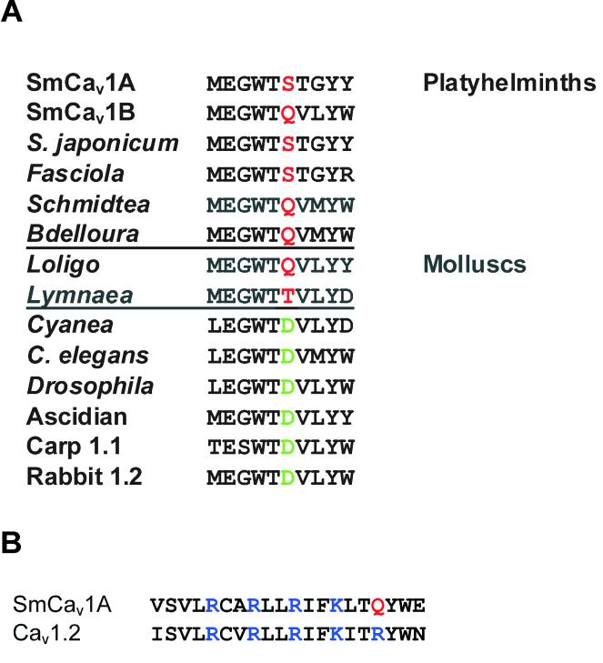
Figure 2. Examples of structural differences between schistosome and other L-type Cav channel α1 subunits.
A. Charge change in the Domain I pore region. The Domain I pore region of L-type α1 subunits is shown. Note the shaded residues four spots C-terminal from the absolutely conserved glutamic acid (E) that forms part of the selectivity gate. In L-type channels from most phyla, this residue is a negatively-charged aspartic acid (D, green). However, in platyhelminths and molluscs, both of which are classed as lophotrochozoans, this residue is uncharged (red). This is interesting, because a non-charged residue at this position is characteristic of some non L-type channels (eg, Cav2.3). Accession numbers for sequences are: SmCav1A, AF361884; SmCav1B, Smp_159990; S. japonicum, Sjp_0099010; Fasciola (F. hepatica), Schmidtea (S. meditteranea), and Bdelloura (B. candida) are from DNA fragments we sequenced; Loligo (L. bleekeri), D86600; Lymnaea (L. stagnalis), AF484081; Cyanea (C. capillata), AAC63050; C. elegans, AAC47755; Drosophila, AAA81883; Ascidian (Halocynthia roretzi), BAA34927; Carp (Cyprinus carpio) Cav1.1, P22316; Rabbit (Oryctolagus cuniculus) Cav1.2, NM_001136522. B. SmCav1A contains one fewer positively charged residue in II-S4. Shown are residues in the fourth transmembrane region of domain II from SmCav1A and rat Cav1.2 (M67515). SmCav1A contains one fewer positively charged residue than Cav1.2. Positively charged arginines (R) and lysines (K) are highlighted in blue, and the substituted glutamine (Q) in SmCav1A is highlighted in red.

