
Figure 8.
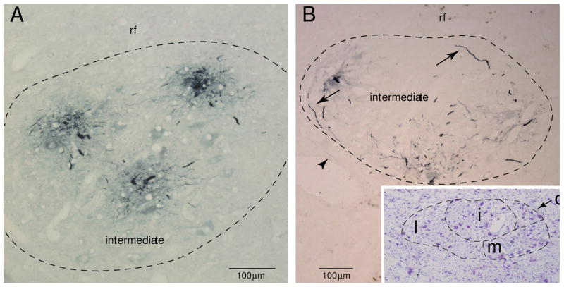
Plate of photomicrographs demonstrating immunohistochemical localization of Lucifer yellow (LY) filled OO motor neurons (stained blue). A: Immunohistochemically localized dendritic branches from 3 filled OO motor neurons in Case 2. In this section, all visible dendritic processes are confined to the intermediate subnucleus. B: LY filled dendrites within the intermediate subnucleus in Case 1. Dendrites located at the periphery of the subnucleus (see arrows) commonly arched to follow the curvature of the boundary. The arrowhead identifies a rare distal dendritic segment within the lateral subnucleus. The inset shows the adjacent tissue section stained with thionin for Nissl substance which aided in defining the external boundary of the facial nucleus and the lateral (l), medial (m), intermediate (i) and dorsomedial (d) subnuclei