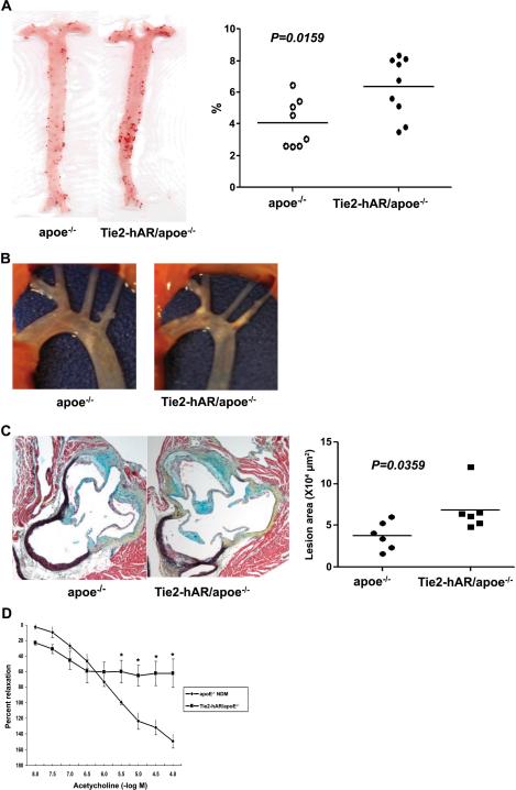
Figure 6.
Whole aorta and aortic root lesions of STZ-induced diabetic apoE−/− and Tie2-hAR/ apoE−/− male mice in early atheroscleorosis. Mice were injected with STZ (50mg/kg mouse body weight) for 5 days to induce diabetes and remained on chow diets for 12 weeks until sacrificed for atherosclerosis analysis. A) Oil red O enface staining in whole aorta and the percentage of oil-red O positive area (n=8–9/each groups) p<0.01. B) Images of aortic arch of STZ-induced diabetic apoE−/− and Tie2-hAR/ apoE−/− male mice. C) The lesion areas of aortic roots with MOVAT staining. Individual lesion positive areas in aortic root cross sections from mice were measured. D) Endothelium-dependent vasorelaxation tested in isolated mouse aortic rings from non-diabetic apoE−/−and Tie2-hAR/apoE−/− (n=4 and 3 respectively), sacrificed at 20 weeks of age. Relaxation is reported as percent of initial phenylephrine precontraction. Comparisons were conducted among groups for each agonist dose. *P < 0.05, apoE−/− versus Tie2-hAR/ apoE−/− (doses >10–5.5 M).

