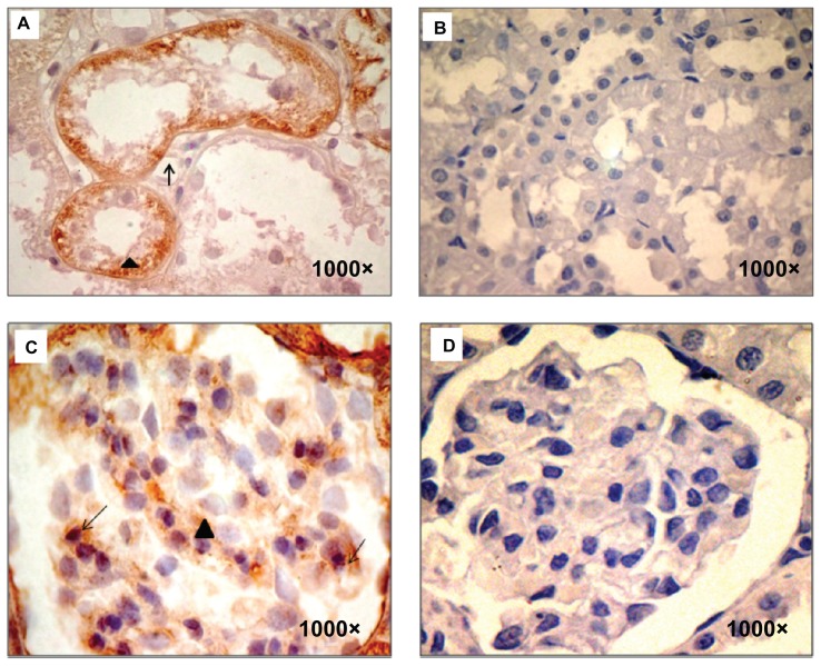
Figure 2.
TGF- β1 immunodetection. (A) sStx2-treated rats showing TGF-β1 expression in the basolateral membrane (black arrow) and the cytoplasm (black arrowhead) of renal tubules, (B and D) control rats showed no stain for TGF-β1, (C) sStx2-treated rats showing expression in the mesangium (black arrowhead) and podocytes (black arrow).
Note: Magnification at 1000×.
Abbreviation: sStx2, supernatant Shiga toxin type 2.

