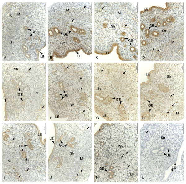
Figure 2.
Localization of EP and FP protein and their regulation by estradiol. Immunohistochemical localization of prostaglandin receptors in luminal epithelium (LE), glandular epithelium (GE), stroma (Str), myometrium (M) and blood vessels (arrows) in the uteri of ovariectomized controls and different doses of estradiol (E2) treated rats. Representative images of EP2 (A-D); OvxC (A), treatment with 1 μg E2 (B), 2.5 μg E2 (C) and 5 μg E2 (D), EP3 (E-H); OvxC (E), treatment with 1 μg E2 (F), 2.5 μg E2 (G) and 5 μg E2 (H). Representative images of EP1 (I), EP4 (J), FP (K) immunostaining and a negative control (L). Magnification - 200X, Scale bar = 100 μm.

