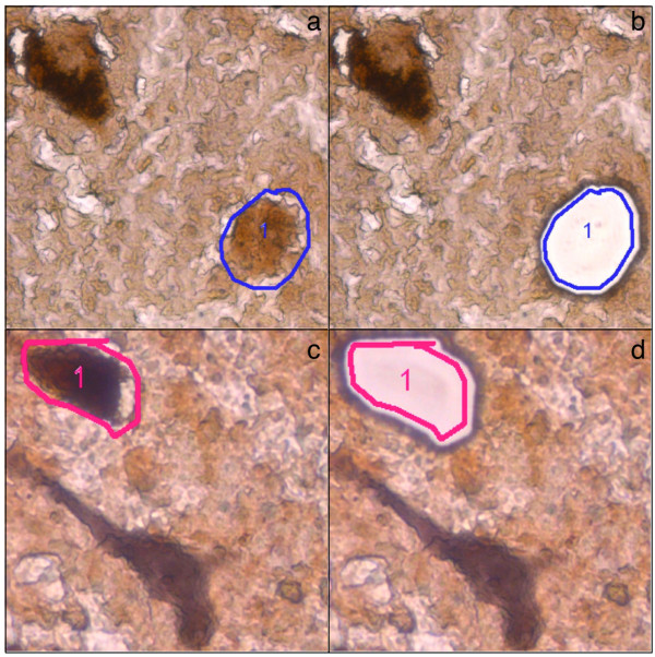
Figure 1.
Immunohistochemical identification of target neurons. (a+b) Pigmented vs. non-pigmented catecholaminergic neurons. TH+ neurons were identified by their brown cytosolic reaction product. Pigmented (TH+/NM+; top left) and non-pigmented (TH+/NM-; bottom right, marked for LMD) neurons were further distinguished by the visible presence or absence of NM. (a) TH+/NM- neuron before and (b) after LMD. (c+d) Catecholaminergic vs. non-catecholaminergic neurons. NeuN+ immunoreactivity results in a grey appearance of TH- neurons due to a DAB/Nickel reaction (bottom neuron). (c) TH+/NM+ neuron before and (d) after LMD.

