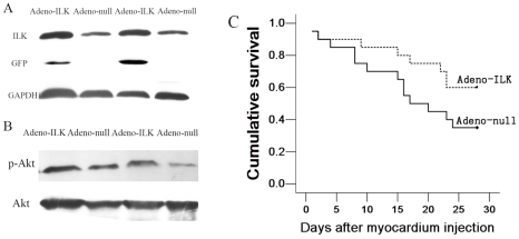
Figure 1. ILK gene therapy increases left ventricular ILK expression and activity, improves survival in doxorubicin-induced cardiomyopathy.
A, Western blot showing elevated ILK protein expression in the left ventricular tissue of the adeno-ILK group as compared with the adeno-null group. The blot also shows green fluorescent protein (GFP) expression in adeno-ILK, but not adeno-null, treated hearts. Glyceraldehyde-3-phosphate dehydrogenase (GAPDH) expression is also shown as a housekeeping protein. B, Western blot showing left ventricular phospho-Akt (pAkt) and Akt expression. The ratio of pAkt to Akt was increased following adeno-ILK delivery relative to adeno-null control, in line with increased Akt phosphorylation due to elevated ILK activity. C, Kaplan-Meier survival curve for doxorubicin-treated rats treated with adeno-ILK (n = 20) and adeno-null (n = 20) (p = 0.093).

