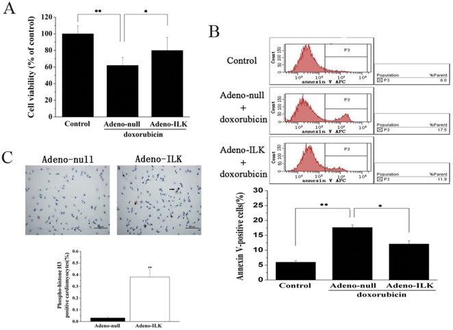
Figure 8. Adeno-ILK prevents apoptosis, and promotes proliferation, of cultured neonatal rat cardiomyocytes treated with doxorubicin.
A, Effect of doxorubicin on cell viability, as assessed by MTT assay, and the effect of ILK on this. n = 6 per group. *, **P<0.05 and <0.01 respectively. B, Effect of doxorubicin on cardiomyocyte apoptosis, as assessed by annexin V expression (determined by flow cytometry), and the effect of ILK on this. Necrotic cells were excluded from the analysis by gating on PI-negative cells. n = 6 per group. *, **P<0.05 and <0.01 respectively. C, Effect of ILK on proliferation of neonatal rat cardiomyocytes treated with doxorubicin, as assessed by phosphohistone-H3 staining (brown); this showed more positive nuclei (arrow) in the adeno-ILK group than the adeno-null group. Graph shows accumulated results of n = 6 experiments. **P<0.01 versus adeno-null group. Scale bar, 100 µm.

