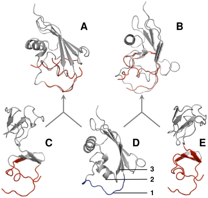
Figure 3. Example of docking based on 12 Å and 16 Å interface libraries.
3sic ligand (gray ribbons in A, B, D) was aligned with fragments of 1oyv ligand (red) extracted using 12 Å (A) and 16 Å (B) interface cutoffs. For comparison, the entire structure of 1oyv ligand is shown with 12 Å (C) and 16 Å (E) fragments (red). The entire structure of 3sic ligand with the loop participating in binding (blue) is shown in D. Binding loop in 3sic ligand is marked 1, and α-helix and β-sheet closest to this loop are marked 2 and 3, respectively.

