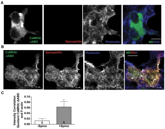
Figure 5. Spinophilin targets CaMKII to F-actin.
A. Compressed Z-stack of confocal images of HEK293 cells expressing GFP-tagged CaMKIIβ ΔABD. F-actin was detected using a far-red phalloidin stain. Scale bar: 10 mM. B. Compressed Z-stack of confocal images of HEK293 cells expressing GFP-tagged CaMKIIβ ΔABD and myc-spinophilin. F-actin was detected using a far-red phalloidin stain. Scale bar: 10 mM. C. Intensity correlation quotient showing significant co-localization of CaMKIIβ with phalloidin in the presence (+Spino), but not absence (−Spino), of spinophilin. Values are expressed as the mean±S.E.M from analyses of the indicated numbers of cells. * P<0.05.

