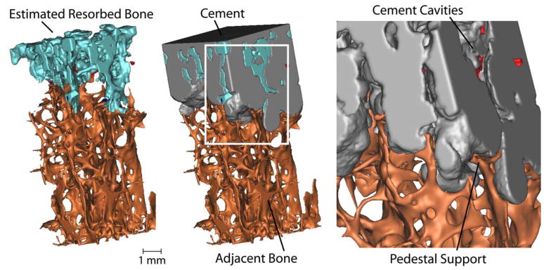
Figure 3.
3-D reconstruction showing estimated resorbed bone following 10 years in service. Cement cavities with small regions of isolated bone (red) are evident in the inset image (right). Following bony remodeling, there appears to be trabecular pedestal support of the cement layer. The specimen was taken from a region under the tibial tray with cross sectional area dimensions of 4 mm × 4mm.

