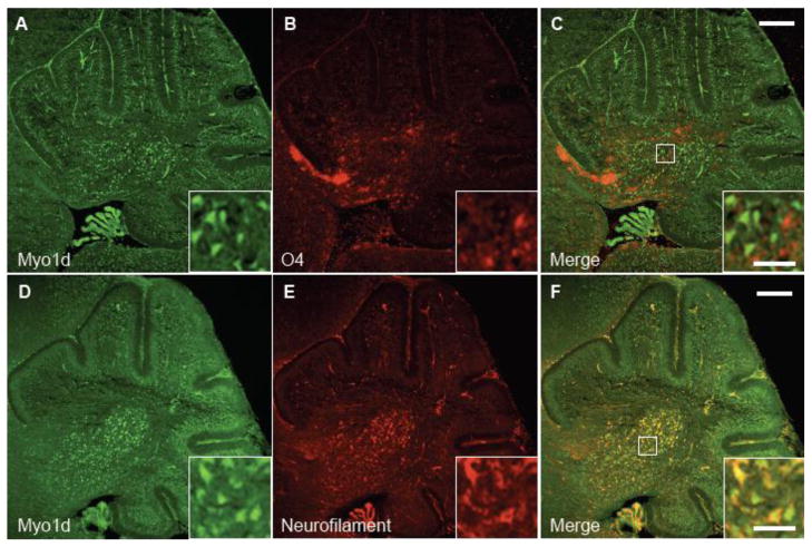
Figure 4. Myo1d is predominantly expressed in neurons at P3.
Mouse cerebella were labeled with antibodies specific for Myo1d (A) and oligodendrocyte progenitor marker, O4 (B). C) Myo1d expression does not overlap with O4. However, when cerebella were stained with antibodies for Myo1d (D) or neurofilament (E), there was greater overlap between both proteins (F). Bar, 200 μm; inset, 50 μm.

