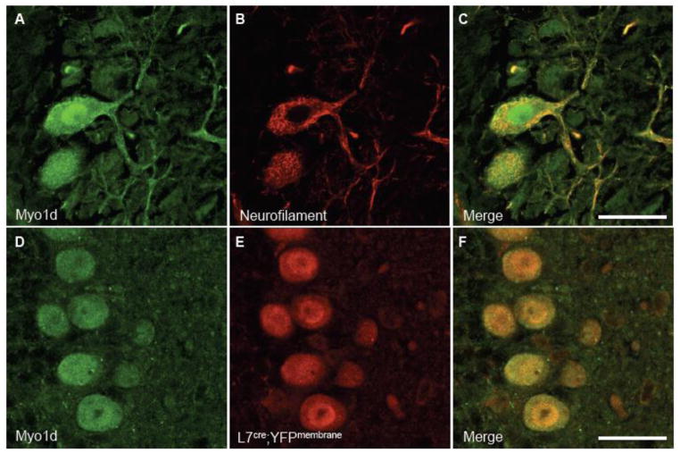
Figure 7. Myo1d exhibits cytosolic and dendritic subcellular localization in Purkinje cells.
We colabeled sagittal cross sections of P14 tissue with antibodies for Myo1d (A) and neurofilament (B). A) Myo1d has punctate cytosolic localization pattern and appears along dendrites. The motor appears more densely along the cell cortex. (D–F) Antibodies targeting Myo1d and GFP were applied to coronal cross sections of L7cre;YFPmembrane mouse cerebella. (D) Myo1d is present in Purkinje cells with L7, a specific Purkinje cell marker. Bar, 25 μm.

