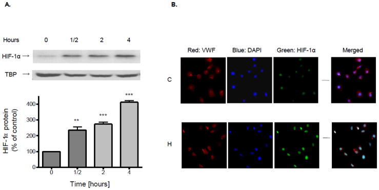
Figure 1.
Confluent brain microvascular endothelial cell cultures were subjected to hypoxia (1% O2) for various periods of time. (A) Nuclear proteins were extracted and resolved using SDS-PAGE. HIF-1α or TBP protein was detected by Western blot probed with corresponding antibodies. Data in the bottom panel are the means ± SD of 3 experiments and expressed as percent of untreated control. **p<0.01, ***p< 0.001 vs. 0 h. (B) After exposure to normoxia (C) or 1% O2 hypoxia (H) for 4 h, brain microvascular endothelial cell cultures were processed for immunofluorescence. Cultures were fixed, incubated with antibodies to VWF and HIF-1α and counter stained with DAPI.

