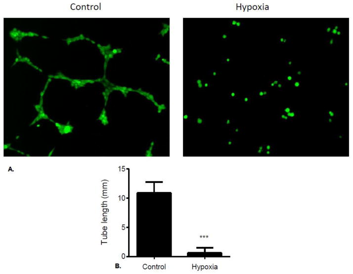
Figure 6.
(A) Brain microvascular endothelial cells were seeded onto the layer of matrix at 105 cells/well, and maintained in DMEM supplemented with 10% FBS. Plates were incubated at 21% O2 (Control) or 1% O2 (Hypoxia) at 37°C for 4 h and then stained with fluorescent dye Calcein for 30 min. Tube-like structures were visualized and captured using Olympus IX71 microscope at 10x magnification. (B) Tube length was analyzed and quantitated using image processing software (ImageJ) available from the National Institutes of Health.

