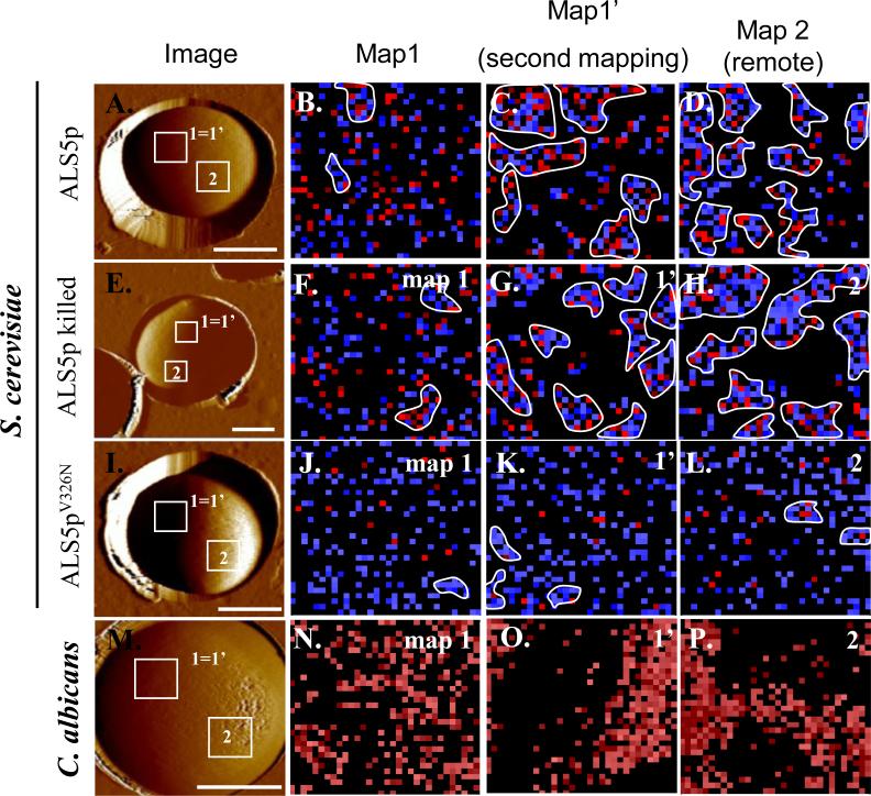
Figure 2.
Induction of surface nanodomains follows application of extending force to individual Als molecules on the surface of yeasts. On the left are AFM images of yeast trapped in a microporous membrane. The second column shows maps of position of Als proteins within the 1 μm square marked “1=1’.” Blue pixels show interactions of <150 pN and red pixels show interactions resistant to forces >150 pN. Clusters of 10 or more contiguous colored pixels are outlined in white [13]. The third column shows clustering of the Als molecules in a second mapping of the same region of the cell walls, and the last column shows similar clustering in a remote region (Map 2). Clustering is a result of surface amyloid formation, as shown by lack of clustering in cells expressing the non-amyloid V326N form of Als5p. Reprinted with permission from Garcia et al. [13].

