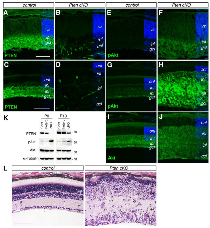
Figure 1. Conditional deletion of Pten enhances Akt phosphorylation and disrupts retinal differentiation.
(A–J) Immunolabeling of control (A, C, E, G, I) and Pten cKO mutant (B, D, F, H, J) retinas at P0 (A, B, E, F) and P18 (C, D, G–J) for PTEN (A–D), pAkt (E–H), and Akt (I, J). Pten cKO retinas show reduced PTEN and increased p-Akt expression.
(K) Western blot analysis of PTEN, p-Akt (Ser473), total Akt, and α-Tubulin in P0 and P13 retinal extracts. MW markers (for 50 kD) are indicated on the right of the panels. (L) H&E staining of control and Pten cKO retinas at P18. Note that the Pten mutant retina shows a thinner outer nuclear layer and significantly expanded inner retina. gcl, ganglion cell layer; inl, inner nuclear layer; ipl, inner plexiform layer; onl, outer nuclear layer; vz, ventricular zone. Scale bars, A for (A, B, E, F), C for (C, D, G–J), L, 100 μm.

