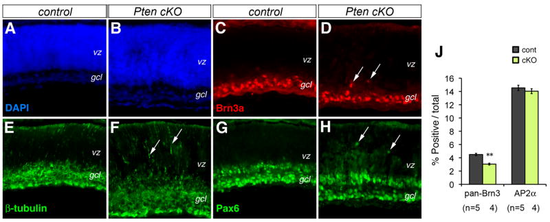
Figure 2. Pten conditional deletion affects retinal cell migration.
(A–H) Immunolabeling of control (A, C, E, G) and Pten cKO (B, D, F, H) retinas at P0 for neuronal markers Brn3a (C, D), β-Tubulin (E, F), and Pax6 (G, H). Arrows point to mislocalized cells.
(J) Quantification of RGCs and amacrine cells by FACS at P0. Percentages of marker-positive cells among total cells are shown (Individual eyes (n) are indicated below the bar graph. Mean ± S.E.M. **, p<0.01).
gcl, ganglion cell layer; vz, ventricular zone. Scale bar, A for (A–H), 100 μm.

