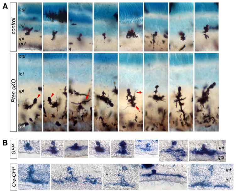
Figure 6. Pten deficiency causes abnormal arborization of amacrine interneurons.
(A) Golgi staining of P45 control (top row) and Pten cKO (bottom row) retinas. Positive stained mutant cells show either Arrowheads point to abnormally positioned cell soma. Arrows indicate overly elaborate dendrites.
(B) Alkaline phosphatase histochemical labeling of individual transfected Ptenfl/fl retinal cells at P21. Ptenfl/fl retinas were electroporated with the control (LIA and GFP, top row) or cre-expressing (LIA, GFP and cre, bottom row) at P0 in vivo. Cre-expressing DNA transfected amacrine cells show more elaborate dendritic morphology.
gcl, ganglion cell layer; inl, inner nuclear layer; ipl, inner plexiform layer; onl, outer nuclear layer. Scale bar, A for (A, B), 40 μm.

