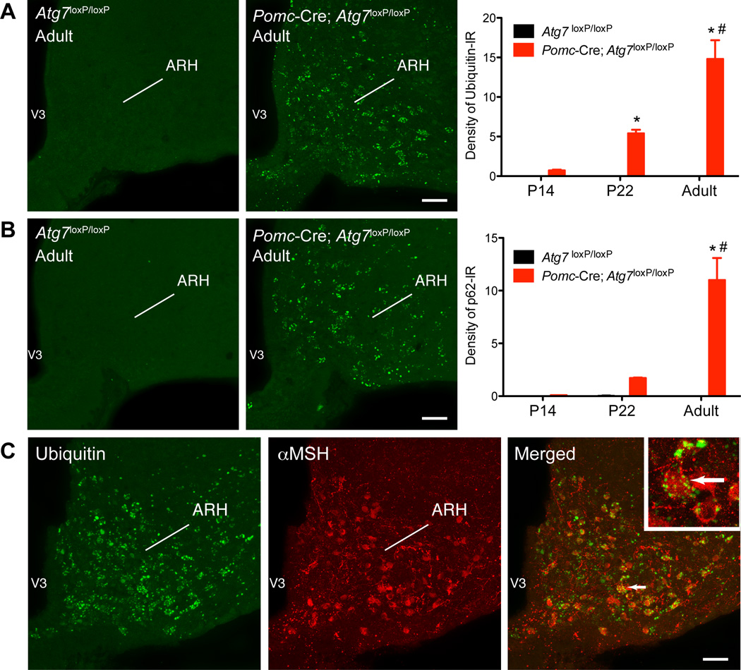
Figure 3.
Lack of autophagy in POMC neurons leads to the gradual accumulation of ubiquitin aggregates in the arcuate nucleus. Quantification of (A) ubiquitin- and (B) p62-immunoreactivity in the arcuate nucleus (ARH) of P14, P22, and adult (15- to 17-week-old) Atg7loxP/loxP (n = 4) and Pomc-Cre; Atg7loxP/loxP (n = 4) male mice. (A, B) Confocal images illustrating (A) ubiquitin- and (B) p62-immunoreactivity in the ARH of adult Atg7loxP/loxP and Pomc-Cre; Atg7loxP/loxP mice. (C) Confocal images showing the presence of ubiquitin-immunoreactivity (green fluorescence) in aMSH-positive cells (red fluorescence) of an adult Pomc-Cre; Atg7loxP/loxP mouse. The arrow points to a doubled labeled cell. V3, third ventricle. Values are shown as mean ± SEM. *P < 0.05 versus P14; #P < 0.05 versus P22. Scale bars, 50 um.

