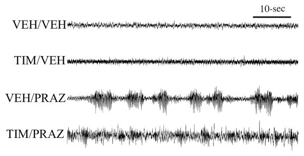
Figure 7.
Synergistic sedative effects of α1- and β-receptor blockade globally within the brain. Shown are the effects of the β-antagonist, timolol (ICV), the α1-antagonist, prazosin (IP) and combined antagonist treatment on cortical EEG in animals exposed to an arousing brightly-lit novel environment. Animals were treated 30-minutes prior to testing with: 1) ICV vehicle + IP saline (VEH/VEH); 2) 150 μg ICV timolol + IP saline (TIM/VEH); 3) ICV vehicle + IP 500 μg/kg prazosin (VEH/PRAZ,), and; 4) combined timolol + prazosin (TIM/PRAZ). In this figure, EEG traces are from the second 5-min epoch of exposure to the novel environment. Vehicle-treated controls displayed behavioral and EEG indices of alert waking throughout most of the recording session. This was reflected in sustained EEG desynchronization (low-amplitude, high-frequency). β-receptor blockade alone (TIM/VEH) had no effects on EEG activity. α1-receptor blockade alone (VEH/PRAZ) increased the frequency and duration of sleep spindles (high-voltage spindles). In contrast to that observed with β-receptor blockade alone, in the presence of α1-receptor blockade, β-receptor blockade produced substantial increases in large-amplitude, slow-wave activity. Power spectral analysis quantified and confirmed these qualitative observations (data not shown; from62).

