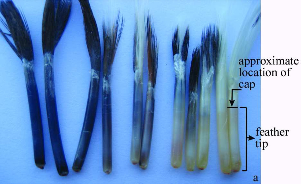
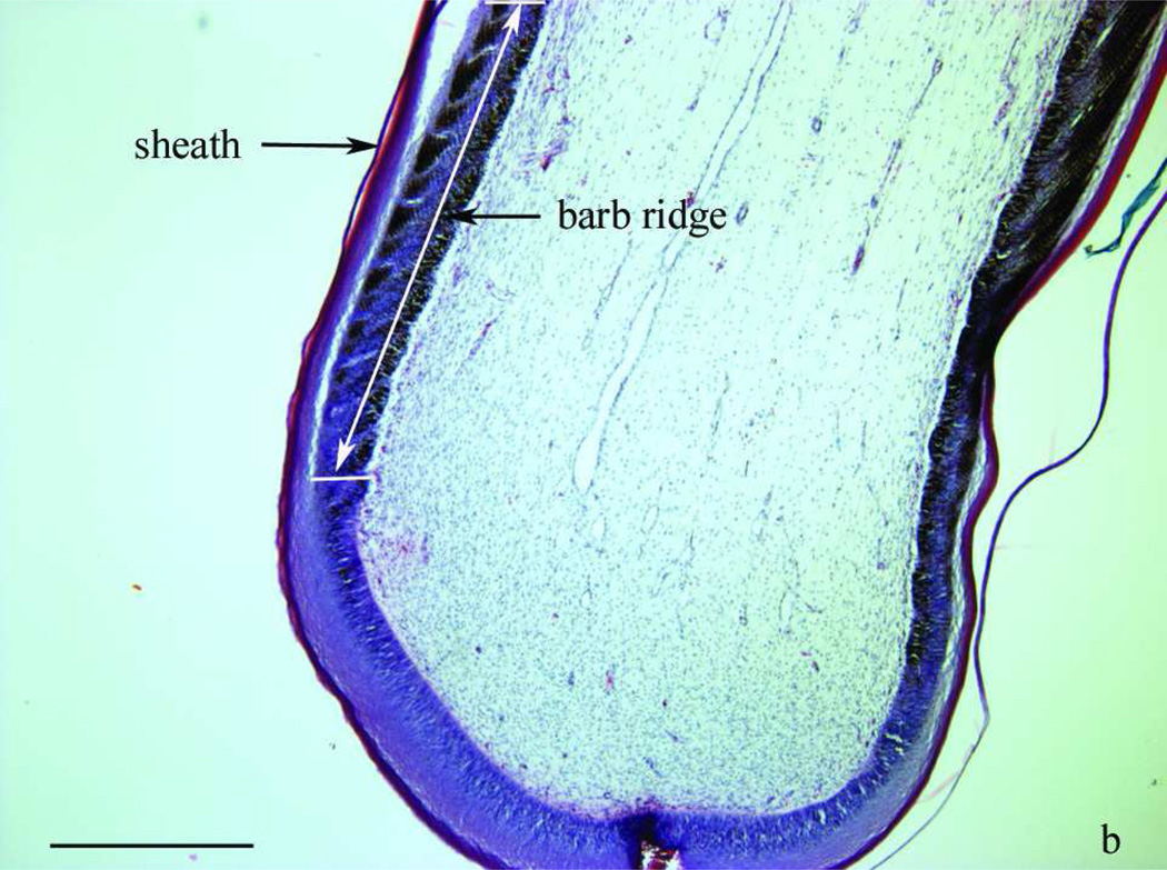
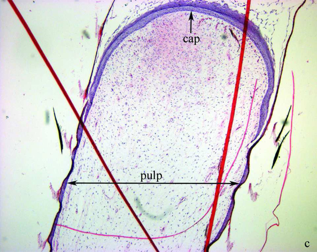
Figure 1.
Morphology of two- to three-week-old growing feathers from SL chickens. a) from left to right: normally pigmented, partially depigmented and completely depigmented growing feathers from SL chickens that developed SLV. Growing feathers can be collected from SLV chickens over the whole course of SLV. The living part of growing feathers (newest growth to the epidermal cap) is referred to as “feather tip”. b) microstructure of the newest growth of a feather tip with normal pigmentation; layers shown from the outside to inside are sheath, barb ridge and pulp. c) a cap formed by epidermal layer enclosing the pulp. Longitudinal sections were stained with H&E stain and examined at 40× (b, c) magnification under a bright field microscope. Bar scale = 1 mm.


